INTRODUCTION
Leishmaniasis is a vector-borne parasitic disease caused by protozoa of the genus Leishmania, transmitted to humans through the bite of infected female sand flies of the genus Phlebotomus (Asia, Africa, and Europe) and Nyssomyia, Migonemyia, Evandromyia Lutzomyia (America) [1-4]. The disease presents in three main clinical forms: cutaneous (CL), mucocutaneous (MCL), and visceral (VL), with CL being the most common form, accounting for the majority of the estimated 600.000 to 1 million new cases reported globally each year [5-7]. Despite this burden, leishmaniasis remains a neglected tropical disease, with significant underreporting cases and diagnostic delay in many endemic areas [8]. Cutaneous leishmaniasis is caused predominantly by species of the Leishmania (Viannia) subgenus, especially L. (V.) braziliensis, L. (V.) guyanensis, and L. (V.) panamensis, as well as the less common L. (Leishmania) amazonensis [7-10].
In Argentina, leishmaniasis is a notifiable disease. Recent official data from 2026 shows a decrease in CL notifications compared with the 2021 - 2025 historical median. However, the temporal trend differs across regions: a decline is observed in the northwest (NOA), while the northeastern region (NEA) remains stable, with increases reported in Chaco and Formosa in recent years. Within the NEA, Chaco and Misiones are among the highest-burden provinces, and L. (V.) braziliensis is the predominant causative agent of CL [10]. Climate change, deforestation, and expanding vector habitats have contributed to the geographic spread of the disease, placing previously low-risk urban populations at risk [7,11].
The classic presentation of CLbegins as a papule at the inoculation site, that typically progresses to a plaque or nodule with a tendency to ulcerate with raised, well-demarcated borders with the classic “volcano crater” appearance [1-3,12]. However, CL can present with a wide spectrum of clinical forms, including papular-nodular, verrucous, and lymphangitic lesions, which may closely mimic other dermatological and oncological conditions[4,8,12-15]. This broad clinical spectrum, together with the frequent absence of amastigotes on routine microbiological and histopathological examination, creates a substantial risk of misdiagnosis. Several have documented CL initially misclassified as cutaneous malignancies, including squamous cell carcinoma, basal cell carcinoma, and, less commonly as soft tissue tumors [13-15].
From a histopathological standpoint, CL lesions usually show a dense dermal inflammatory infiltrate composed of lymphocytes, macrophages, and plasma cells, with variable granuloma formation[3,12,16,17]. In cases with a prominent spindle cell component and reactive fibrohistiocytic proliferation, the morphological overlap with low-grade soft tissue sarcomas, particularly low-grade myofibroblastic sarcoma, can represent a major diagnostic pitfall, especially in laboratories with limited access to molecular diagnostic tools[2,18,19].
We report a case of CL in a young man from Resistencia, Chaco, Argentina, whose initial presentation with a progressive ulcerating skin lesion and a right inguinal mass led to a histopathological diagnosis of low-grade myofibroblastic sarcoma. The definitive diagnosis was ultimately established by real-time PCR for Leishmania spp., and subsequent pathological review reclassified the lesion as a fibrohistiocytic inflammatory process. This case highlights the critical importance of molecular diagnosis in endemic areas and the need to include CL in the differential diagnosis of soft tissue tumors.
CASE PRESENTATION
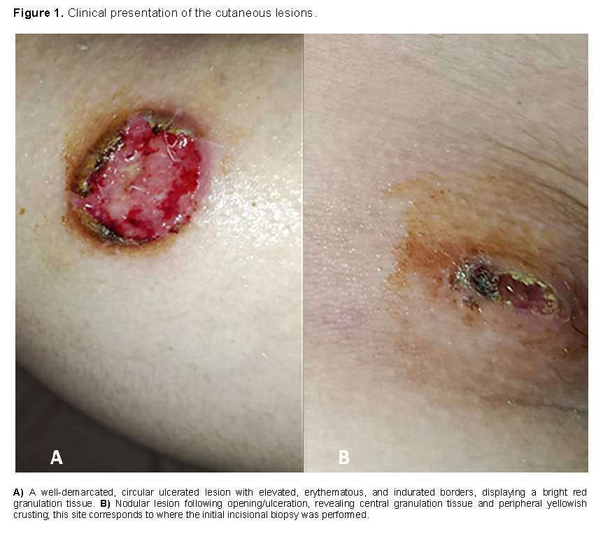
A 19-year-old male from Resistencia, Chaco, Argentina, presented in January 2026 with progressive, ulcerated skin lesion on the right flank concomitant with a painful right inguinal mass that subsequently ulcerated.
The cutaneous lesion began as a small papule at the right flank that enlarged progressively over the following days. A few days later, the patient developed a nodular lesion at the right inguinal region, initially described as furuncle-like. At his first medical consultation, right inguinal inflammation was noted, and an ultrasound, chest X-ray and blood tests were requested. The ultrasound was initially interpreted as an inguinal hernia; however, subsequent surgical assessment suggested enlarged lymph nodes with fluid collection. The patient was then hospitalized for surgical exploration, and an incisional biopsy of the nodular lesion was obtained (Figure 1).
Histopathological examination of the biopsy revealed a low-grade spindle cell neoplasm composed predominantly of fascicles of spindle-shaped cells with eosinophilic cytoplasm and fusiform to oval nuclei. Mitotic activity reached up to 5 mitoses per 10 high-power fields, and no areas of tumor necrosis were identified. Based on these findings, immunohistochemical analysis was recommended. Initial laboratory tests were within normal limits, except for a mildly elevated C-reactive protein (CRP) level (18mg/l). Serologic testing for HIV and VDRL was non-reactive.
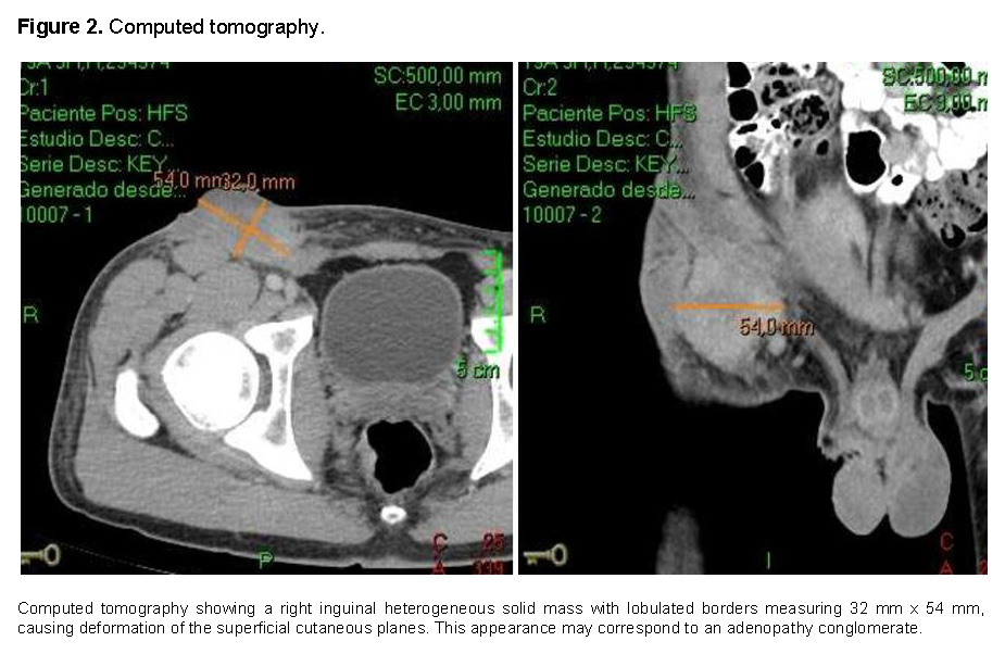
A contrast-enhanced whole-body computed tomography scan showed a heterogeneous solid right inguinal mass measuring 32 × 54 mm with lobulated bordersand deformation of the superficial cutaneous planes, consistent with an adenopathic conglomerate. No visceral involvement, deep lymphadenopathy, or metastases were identified (Figure 2).
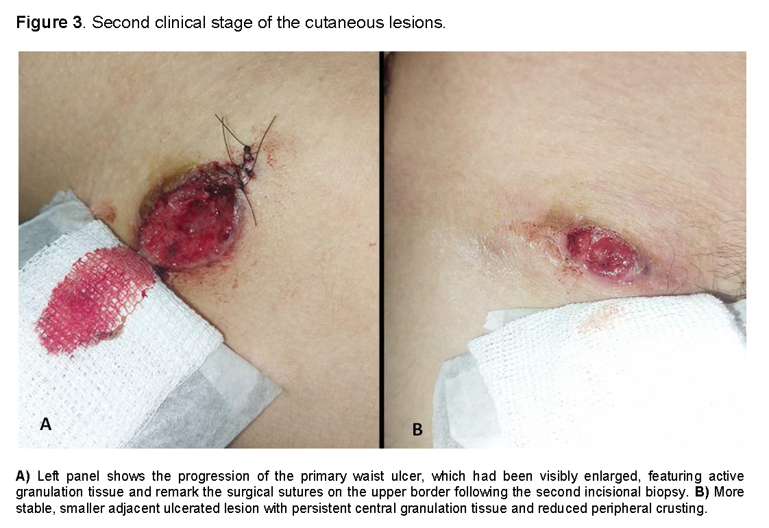
Because of progressive clinical worsening and enlargement of the flank ulcer, a second biopsy of the lesion was performed (Figure 3). Histopathology showed a nonspecific ulcer associated and a mixed inflammatory infiltrate composed of lymphoplasmacytic cells, eosinophils, and macrophages and multinucleated giant cells. Special stains, including PAS and Giemsa were negative. Mycological studies on both a skin lesion scraping and ulcer biopsy did not show fungal or parasitic elements on direct examination and stains, and cultures were negative for fungi, bacteria, and mycobacteria.
Immunohistochemistry results obtained one month later indicated a low-grade myofibroblastic sarcoma in the right inguinal lesion, based on morphological characteristics and immunoprofile (vimentin positive, smooth muscle actin positive, desmin negative, Ki-67 16-18%). The patient was therefore referred to Fleming Hospital in Buenos Aires, for specialized oncological evaluation.
In parallel, a fragment of the second biopsy specimen was sent to the Molecular Biology Laboratory of Instituto de Medicina Regional - Universidad Nacional del Nordeste for real-time PCR tests for Leishmania typing. DNA extraction was performed using the CTAB method [20], and real-time PCR coupled with melting curve analysis was carried out using primers targeting the polymerase II gene and EvaGreen fluorophore [21].The DNA polymerase II gene was selected because it provides species-discriminatory sequence variation and may reduce some of the interpretive limitations associated with multicopy targets [22].
Using the reported sequences for the DNA polymerase II gene, it was determined that, in the region between nucleotides 536 and 649, there were differences in the base composition sequences between different species of this parasite.
The sequences of the primers used were: FW: AGGAGGATGGCAAGCGGAAG and RV: GCGACGGGTACAGGGAGTTG.
The thermal profile used consisted of an initial denaturation cycle at 95°C for 5 min, followed by 40 cycles of denaturation at 95°C for 10 seconds, annealing at 57°C for 40 seconds (s), and extension at 72°C for 10 s. High-resolution melting (HRM) analysis was performed between 78°C and 90°C, increasing the temperature by 0.1°C every 2 s to denature the amplicons, with fluorescence readings taken at each temperature increment.
The PCR-HRM technique allowed obtaining high-resolution average denaturation temperature curves, clearly differentiated and highly reproducible for Leishmania braziliensis and the two samples included as controls (L. infantum, L. panamensis) (Figure 4).
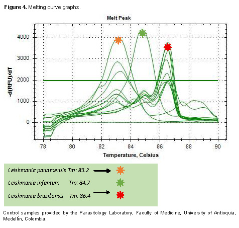
The assay showed a detectable amplification curve, confirming the presence of Leishmania (Viannia) braziliensis. These results were subsequently confirmed using specific PCR [20] and HSP70 PCR, followed by RFLP with Sau3AI [23].
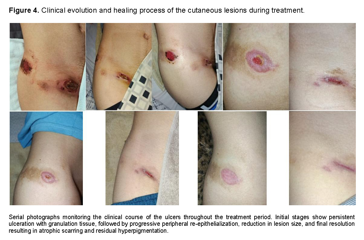
After confirmation of the diagnosis, the patient was evaluated by an infectious diseases specialist and started on treatment with meglumine antimoniate (Glucantime®) at the standard regimen for cutaneous leishmaniasis, intramuscular injections two per day of 1.5 g/5mL solution (41 doses during 21 days). During treatment, serial clinical examinations documented progressive improvement in the skin lesion, with reduction in ulcer size and granulation tissue and gradual re-epithelialization (Figure 5).
On-treatment electrocardiographic monitoring was normal, showing only minor repolarization changes within physiological limits for age. After initiation of meglumine antimoniate therapy, follow-up laboratory tests showed a mild decrease a mild decrease in hemoglobin to 12.00 g/dL and hematocrit to 35.3%, together with a slight increase in hepatic transaminases (AST 42 U/L, ALT 58 U/L). These changes were consistent with the known reversible hematologic and hepatotoxic adverse effects of pentavalent antimonials. White blood cell counts, renal function, and glucose levels remained within reference ranges throughout treatment. The patient completed therapy on April 30, and had an excellent overall clinical outcome.
Two months later, expert review of the paraffin block from the original inguinal biopsy showed a fibrohistiocytic proliferation with fusiform fibroblast-like elements, marked reactive changes, numerous histiocytes containing small rounded intracytoplasmic structures potentially consistent with microorganisms, and chronic active inflammatory infiltrate. The final pathological diagnosis was revised to fibrohistiocytic inflammatory process. This reclassification, in conjunction with the positive real-time PCR result, was considered diagnostic of cutaneous leishmaniasis.
DISCUSSION
Cutaneous leishmaniasis (CL) is consistently reported in northeastern Argentina, with Chaco recognized as one of the highest-burden regions in the country. The two primary vectors responsible for CL transmission are Nyssomyia neivai (a proven vector of Leishmania braziliensis) alongside Migonemyia migonei and the Evandromyia cortelezzii-sallesi complex. Meanwhile, Lutzomyia longipalpis remains the main vector responsible for visceral leishmaniasis (VL) transmission [10,11,24]. The species identified in this case was Leishmania (Viannia) braziliensis, the predominant causative agent of CL in the NEA.
The initial differential diagnosis in our patient included, lymphoma, tuberculous lymphadenitis, endemic mycoses (such as sporotrichosis, paracoccidioidomicosis and chromoblastomycosis), pyogenic abscess, and reactive lymphadenopathy [4]. These possibilities were considered less likely on the basis of negative microbiological cultures, absence of deep lymphadenopathy or visceral involvement on contrast-enhanced CT, and the subsequent clinical and molecular findings.Low-grade myofibroblastic sarcoma (LGMS) is a rare mesenchymal tumor characterized histologically by a fascicular proliferation of spindle cells with myofibroblastic differentiation, low mitotic activity, and the absence of necrosis—features that were initially present in the biopsy of this patient [19]. The immunohistochemical profile of LGMS typically includes vimentin and smooth muscle actin positivity, with a low Ki-67 proliferative index (which was 16-18% in this case), all consistent with the diagnosis presented [2,19]. However, other reactive inflammatory conditions, including leishmaniasis, can generate fibrohistiocytic spindle cell responses that mimic this entity [2,16].
The histopathological diagnosis of CL can be challenging,particularly in cases with a low parasite burden, when amastigotes are not visualized on routine hematoxylin-eosin or Giemsa staining, when there is a prominent inflammatory or fibrohistiocytic reaction, or there are atypical clinical presentations [15-17]. The hallmark of CL histopathology is a dermal lymphohistioplasmacytic inflammatory infiltrate, with variable granuloma formation and, in classic cases, visible amastigotes within macrophages [2]. However, the spectrum of histological patterns is wide, ranging from diffuse macrophagic infiltrates with abundant parasites to well-formed epithelioid granulomas with few or absent organisms [16,25]. In the latter scenario, the reactive spindle cell and fibrohistiocytic components can be mistaken for a mesenchymal neoplasm, as occurred in this case.
Several case reports and series have documented CL initially misdiagnosed as malignancies, including squamous cell carcinoma, epithelial skin cancer, and soft tissue tumors[5,13-15]. The misdiagnosis carries serious consequences: inappropriate surgical procedures, delays in antiparasitic treatment, unnecessary oncologic evaluation, and significant psychological distress for the patient and relatives, all of which were experienced in this case.
The immunohistochemical panel in this case (vimentin positive, SMA positive, desmin negative, Ki-67 16-18%) was consistent with low-grade myofibroblastic sarcoma, but also compatible with a reactive inflammatory process in reactive myofibroblastic lesions[26]. Immunohistochemistry, while valuable in characterizing spindle cell lesions, should always be interpreted in conjunction with clinical and epidemiological data [19]. The importance of requesting molecular confirmation in diagnostically ambiguous cases from endemic regions cannot be overstated [4,16].
Real-time PCR targeting conserved Leishmania genes—including kinetoplast DNA (kDNA), the internal transcribed spacer (ITS), and the RNA polymerase II gene used in this case—has emerged as the most sensitive and specific method for confirming CL, particularly in cases with low parasite burden or atypical histology [1,2,4,27].
High-resolution melting (HRM) analysis, as applied in this case, offers the additional advantage of potential species discrimination without additional processing after the PCR reaction, making it a robust tool in reference laboratories[28]. In this case, the result obtained was decisive in redirecting the clinical management from oncological to antiparasitic treatment.
The negativity of Giemsa staining in both the skin scraping, the biopsy specimen, and the expert review section in this case is consistent with published data on the diagnostic sensitivity of direct microscopy in CL. Sensitivity of Giemsa-stained and histological samples ranges from 15% to 70%, depending on lesion chronicity, parasite burden, and sample quality, with the lowest yields observed in chronic ulcerated lesions and in infections caused by L. (V.) braziliensis, where the inflammatory response tends to suppress parasite replication [4,16,27,29]. The histopathological finding of small rounded intracytoplasmic bodies within histiocytes — morphologically suspicious for amastigotes but not confirmed by Giemsa in this case— exemplifies the diagnostic uncertainty that requires molecular confirmation. Real-time PCR with high-resolution melting analysis, as performed in this case, overcomes this limitation with a reported sensitivity of 85-98% in skin samples, even in chronic lesions [23,27,29].
Meglumine antimoniate (Glucantime® 1.5g/5mL), a pentavalent antimonial compound, remains the first-line treatment for CL in Argentina and throughout Latin America, in accordance with the national guidelines of the Argentine Ministry of Health and PAHO recommendations[7-9]. The standard regimen for CLis 20 mg/kg/day for 20 to 30 days, typically administered intramuscularly. In this patient, 42 injections were administered over 21 days, with excellent clinical response. The mild anemia (hemoglobin decreased from 13.60 to 12.00 g/dL) and mildly elevated liver transaminase levels (AST 42 U/L, ALT 58 U/L) observed during treatment are well-recognized, reversible adverse effects of pentavalent antimonials, reflecting hematologic and hepatotoxicity that typically resolves upon completion of treatment.
Cardiac monitoring was appropriately performed (ECG normal, with minor repolarization changes within physiological limits for age), given the known risk of QTc prolongation associated with antimonial therapy [30].
This case demonstrates that CL can present with histopathological features closely mimicking low-grade soft tissue sarcoma, leading to potentially harmful diagnostic and therapeutic delays. In endemic areas such as Chaco, Argentina, CL should be systematically included in the differential diagnosis of any soft tissue mass or tumor, regardless of initial biopsy findings. Negative Giemsa staining in chronic CL, particularly when caused by L. (V.) braziliensis, does not exclude CL. Real-time PCR is the gold standard confirmatory method when conventional studies are inconclusive, with high sensitivity even when conventional microbiological and histopathological studies are inconclusive.
Early and accurate diagnosis is essential to initiate appropriate antiparasitic therapy, avoid unnecessary surgical or oncological interventions, and prevent long-term morbidity. This case also underscores the need for multidisciplinary collaboration—integrating infectious disease, dermatology, pathology, and molecular biology—in the management of atypical CL presentations. Treatment response is excellent when appropriate antiparasitic therapy is initiated, even after diagnostic delay.