CASE REPORT
This is a 19-year-old male patient, the only child of non-consanguineous parents, whose pregnancy was monitored from the beginning, with threatened preterm labor occurring at 26 weeks. Ultrasound examinations reported a fetus small for gestational age. Delivery was by cesarean section due to acute fetal distress at 35 weeks, with a birth weight of 1.8 kg, length of 40 cm, and head circumference of 28.3 cm, with an Apgar score of 8/9.
He presented with neonatal hyperbilirubinemia and was diagnosed with cardiac abnormalities consisting of a patent ductus arteriosus that closed shortly thereafter, pulmonary valve stenosis with a gradient of 27 mm, a high muscular ventricular septal defect (VSD) with an approximately 4-mm left-to-right shunt, and mild stenosis of the left pulmonary artery branch.
Microcephaly was detected at 10 months of age, together with global developmental delay. At 2 years and 11 months, a blood amino acid profile was performed and was normal, and a karyotype revealed 45,X,del(8)(p22)[43]/46,XY[57]dn. These abnormalities were de novo, as the karyotypes of both parents were normal.
Subsequent psychometric evaluations diagnosed moderate intellectual disability with severe language delay, distractibility, and sensory integration dysfunction. At 4 years of age, he underwent surgery to correct alternating strabismus of both eyes, predominantly affecting the right eye, and at 6 years of age, spontaneous closure of the VSD was confirmed.
At 8 years of age, mild thoracic scoliosis was detected, and beginning at 17 years of age, he developed tonic-clonic seizures, currently managed with two medications. Brain MRI detected right mesial temporal sclerosis, which had not been present in several previous studies.
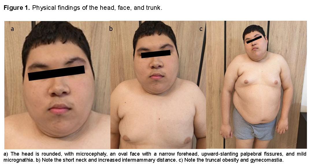
The patient had been lost to Genetics follow-up since childhood and recently returned for reevaluation. On physical examination, our patient has a height of 1.43 m, shorter than the heights of his parents, who measure 1.67 m and 1.56 m. Microcephaly is observed, with a rounded head and narrow forehead.
The face is oval, with low-set ears; the upward-slanting palpebral fissures are narrow, and epicanthal folds are present. The neck is short, with a low posterior hairline. The patient also presents with truncal obesity, increased intermammary distance, and gynecomastia (Figure 1a-c).
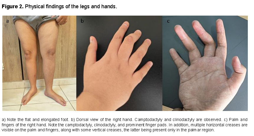
In the upper extremities, the hands have tapered fingers with clinodactyly of the second and third fingers of the left hand and the first, third, and fourth fingers of the right hand. In the lower extremities, the feet are flat, elongated, and slender (Figure 2a-c).
Regarding self-care and hygiene activities such as grooming and toileting, our patient can perform them with partial independence but requires assistance to complete them successfully. Likewise, although he chooses his clothing, he requires assistance with dressing.
Regarding feeding, he eats orally, selects what he wishes to eat, requires assistance serving his food, and eats independently using only a spoon. His diet is widely varied.
Regarding mobility, although he had delayed psychomotor development, he is currently autonomous and independent.
Regarding sensory processing, he was diagnosed with a sensory processing disorder in early childhood. He currently dislikes hot food and exhibits anger and low tolerance toward certain sounds, particularly very loud noises and high-pitched tones.
Regarding communication, his verbal language is almost absent, with a vocabulary of fewer than five words. He usually communicates through gestures to indicate preferences and has developed an alternative and augmentative communication (AAC) method, remarkably using scenes or phrases from audiovisual programs on his mobile device that he knows how to locate and then plays for his caregivers to communicate his meaning.
Regarding his mental health and behavior, he is usually calm and cheerful but becomes irritated when caregivers set limits or in response to certain environmental situations, such as particular sounds, as previously mentioned.
Cognitively, he has moderate-to-severe intellectual disability. He is able to follow instructions, demonstrates considerable ease in using audiovisual media, and has a very notable circumstantial memory for events, people, or images from programs, animated series, and soap operas.
To complement the information obtained from his previous karyotype performed 16 years earlier, and because of the parents’ interest in assessing the risk of other genetic conditions present in the family, molecular testing was performed to delineate the extent of the deletion involving the short arm of chromosome 8 and to identify other potentially affected genes.
A new karyotype was also ordered to corroborate the findings detected in the initial examination.
In addition, the patient was referred to Urology for evaluation because of the risks associated with gonadal dysgenesis in these cases. Testicular ultrasonography revealed a small cyst in the left epididymis and a mild left varicocele, with no other abnormalities.
The patient is followed by Neurology for his diagnosis of refractory epilepsy, for which he receives two anticonvulsants. He is also being followed and evaluated by Endocrinology and Gastroenterology for longstanding dyspepsia. A recent abdominal ultrasound revealed diffuse hepatic steatosis and pancreatic lipomatosis.
Hormonal testing revealed hypogonadism, with low testosterone and free testosterone levels and normal FSH and LH levels.
MOLECULAR TESTING AND KARYOTYPE
MOx 1.0 Multiomic Whole-Genome Analysis (CentoGenome® MOx 1.0)
Genomic DNA was enzymatically fragmented and tagged with Illumina-compatible adapter sequences. Libraries were sequenced from both ends (paired-end) on an Illumina platform to obtain a mean read depth of approximately 30×. A bioinformatic procedure based on Illumina's DRAGEN algorithm, together with CENTOGENE's internal bioinformatic pipeline, was used. Reads were aligned to the Genome Reference Consortium Human Build 37 reference genome (GRCh37/hg19), as well as to the revised Cambridge Reference Sequence (rCRS) of human mitochondrial DNA (NC_012920).
Sequence variants (SNVs/indels) and copy-number variants (CNVs) were identified using DRAGEN, Manta, and internal algorithms. All variants with a minor allele frequency (MAF) below 1% according to the gnomAD database, as well as disease-associated variants described in HGMD®, ClinVar, or CENTOGENE's internal Biodatabank, were analyzed. Although the evaluation focused on coding exons and flanking intronic regions, the entire gene was analyzed for candidate variants potentially associated with the phenotype.
All potential inheritance patterns were considered. In addition, family history and the clinical information provided were considered when assessing the identified variants for pathogenicity and disease causality. Sequence variants were classified into five classes (pathogenic, likely pathogenic, variant of uncertain significance [VUS], likely benign, and benign) according to ACMG/AMP guidelines together with ClinGen recommendations. Copy-number variants (CNVs) were classified into five classes (pathogenic, likely pathogenic, variant of uncertain significance [VUS], likely benign, and benign) according to ACMG/ClinGen guidelines for CNV classification.
Other clinically relevant variants could also be detected (e.g., risk factors or modifier variants). All relevant variants related to the patient's phenotype were reported. Benign and likely benign variants were not reported. For CentoGenome® MOx, when biochemical analysis is applicable, it is performed following the detection of relevant variants by sequencing specific genes. This improves the diagnosis of metabolic disorders, optimizes variant classification, and helps determine their ultimate contribution to the phenotype. The list of available enzyme activity assays and biomarkers can be consulted at www.centogene.com/mox.
CNVs of uncertain significance with no apparent relationship to the patient's phenotype are not reported. All mitochondrial variants with heteroplasmy levels of 15% or higher are reported. For the detection of SNVs and indels in regions selected for subsequent analysis, a sensitivity of 99.9%, specificity of 99.9%, and accuracy of 99.9% were achieved. The CNV detection software has a sensitivity greater than 95%.
CENTOGENE has established strict quality criteria and validation processes for variants detected by NGS. Relevant variants detected by NGS that do not meet internal quality and validation standards—including, but not limited to, variants with unclear heterozygosity, including mosaic variants, CNVs spanning complex genomic regions, and CNVs smaller than 500 kb—are confirmed using orthogonal methods.
CNVs involving clinically significant imprinted regions were additionally investigated using MS-MLPA analysis. Consequently, a specificity >99.9% was ensured for all reported variants.
Repeat expansion screening was performed for the following genes: AR, ATN1, ATXN1, ATXN2, ATXN3, ATXN7, ATXN8OS, ATXN10, CACNA1A, CNBP, CSTB, C9orf72, DMPK, FMR1, FXN, HTT, JPH3, NOP56, PABPN1, PHOX2B, PPP2R2B, PRNP, and TBP. Technical results from repeat expansion screening were correlated with the clinical information provided. Any identified repeat expansion considered relevant to the phenotype was confirmed using an orthogonal method.
GBA1 screening was performed using the Gauchian algorithm to detect recombination events affecting the region spanning exons 9–11 (NM_000157.3), a region homologous to GBAP1. Any detected recombination event would be reported only if considered relevant to the phenotype.
Spinal muscular atrophy (SMA) screening was performed using the SMN Caller algorithm to determine the SMN1 gene copy number. Any detected CNV would be confirmed using an orthogonal method and reported only if considered relevant to the phenotype.
Screening for uniparental isodisomy (isoUPD) was performed using an internal algorithm to detect Mendelian inheritance abnormalities and identify regions of homozygosity (ROH) in known clinically relevant chromosomal regions (6q24, 7, 11p15.5, 14q32, 15q11q13, 20q13, and 20).
Karyotype
Three milliliters of blood were obtained from the patient by venipuncture and placed in a sterile sodium heparin tube maintained under vacuum to prevent bacterial contamination. A total of 0.5 mL of peripheral blood was added to each tube and mixed by inversion. The tubes were incubated at 37°C for 72 hours, after which the samples were processed by adding 100 µL of colcemid to each tube and mixing by inversion, followed by incubation at 37°C for 30 minutes.
The tubes were then centrifuged at 2000 rpm for 10 minutes. The supernatant was discarded, leaving approximately 1 mL for resuspension of the cell pellet at the bottom of the tube using a transfer pipette. After vortexing, 5 mL of KCl at 37°C was slowly added while vortexing, followed by incubation for 30 minutes at 37°C.
Subsequently, 1 mL of fixative solution was added, followed by incubation for 10 minutes at room temperature and centrifugation at 2000 rpm for 10 minutes. The solution was resuspended and slides were prepared. The slides were flame-treated over a burner. They were allowed to dehydrate for at least one day before banding. Fifty metaphases were examined and photographed.
RESULTS
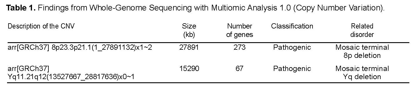
NGS-based CNV analysis identified an approximately 27.9-Mb terminal loss (copy number: 1–2; classification: pathogenic) involving chromosomal cytobands 8p23.3p21.1. This finding was confirmed by chromosomal microarray analysis (CMA). CMA data suggest mosaicism for the detected chromosomal abnormality, with an estimated mosaicism level of approximately 88.6%. This result is compatible with a genetic diagnosis of mosaic terminal 8p deletion.
Additionally, an approximately 15.3-Mb loss (copy number: 0–1; classification: pathogenic) encompassing the entire long arm of chromosome Y, including the AZF regions, was identified. This finding was confirmed by CMA. CMA data suggest mosaicism for the detected chromosomal abnormality, with an estimated mosaicism level of approximately 69.7%. Terminal deletions removing all or part of the AZF regions together with the terminal q12 band of chromosome Y are often associated with impaired spermatogenesis. (See Table 1 and Supplementary Table 1.)
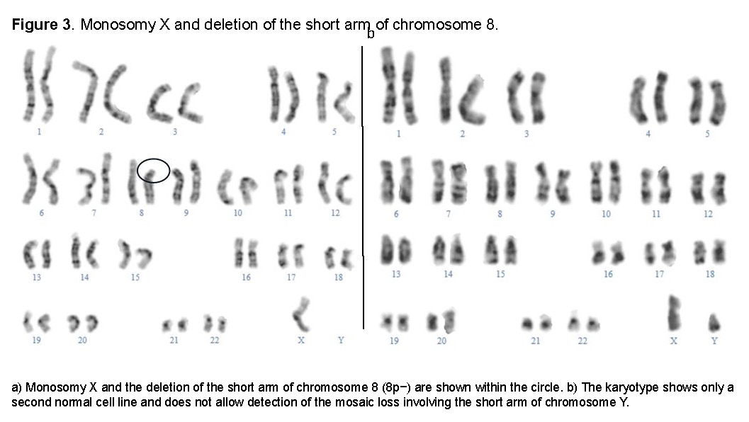
The karyotype (see Figure 3) was reported as:
mos45,X,del(8)(p21-pter)[27]/46,XY[23].
DISCUSSION
In the process of understanding the genome and its function, the identification of complex genetic abnormalities always represents an interesting finding in which the events of permanent change that normally occur in nature become more tangible. In our patient, both structural and numerical de novo chromosomal changes coexist.
We could begin by analyzing the phenotypic features as a unique and unpredictable cumulative effect of three mosaic genetic abnormalities, each having its own individual effect, and examine how each may have contributed to producing the resulting clinical presentation. In this regard, the karyotype and molecular study provide complementary perspectives. The molecular study reveals abnormalities invisible to the karyotype but, because of its limitations, fails to identify that one of the cell lines contains both monosomy X and the 8p deletion.
In this case, the patient would have three cell lines and three chromosomal abnormalities: one aneuploidy and two structural abnormalities. There is one normal cell line and two abnormal cell lines that could be designated as 45,X,del(8)(p21.1-p23.3)/46,Xdel(Y)(q11.21-q12)/46,XYdn when all the information provided by both the molecular studies and the karyotypes is integrated. For practical purposes, the two karyotypes are concordant in their findings, considering that they were performed 16 years apart.
Many patients have been reported with rearrangements involving the short arm of chromosome 8, including inversions/duplications/deletions [invdupdel(8p)], duplications (dup8p), deletions (del8p), and ring chromosome 8p [1,2,3]. More recent publications have focused on detailed genotype-phenotype correlations to establish management guidelines [4] and on the development of new therapies [5], with support from foundations dedicated to this condition, such as Project 8p Foundation (http://www.project8p.org).
A significant proportion of our patient's clinical manifestations correspond to those associated with the 8p terminal deletion, or 8p− syndrome, including microcephaly, congenital heart defects, intellectual disability, recurrent infections in early childhood, obesity, short stature, and dysmorphic features such as epicanthal folds, strabismus, upward-slanting palpebral fissures, rotated low-set ears, a short neck, and hands with tapered fingers, as described previously [6,7,8,9]. This is expected given the high degree of 8p− mosaicism, approximately 90%.
A comprehensive analysis of the 273 genes reported in this case as being involved in the 27.9-Mb deletion helps us understand the phenotype, as analyzed by Okur et al. [2]. Although the vast majority of the genes listed are non-coding RNAs, pseudogenes, uncharacterized loci, or genes exclusively associated with recessive conditions [10], several are already clearly linked to clinical manifestations, such as GATA4 and SOX7, which are involved in congenital cardiac malformations [11,12], with the latter also associated with autism and intellectual disability [13]. GATA4 has also been associated with gonadal dysgenesis [14]. A significant number of other genes in this region are associated with neurodevelopment and behavioral disorders [11,15]. Abbott et al. (2026) performed a comprehensive analysis correlating epilepsy with 8p-related disorders [16].
The high incidence of deletions and rearrangements involving 8p may arise through non-allelic homologous recombination (NAHR) due to the presence of clusters of olfactory receptor (OR) and defensin genes, which contain highly repetitive and nearly identical DNA sequences. During maternal meiosis, this makes the region highly susceptible to errors during recombination processes [17].
Regarding the male phenotypic characteristics and fertility of our patient, these would be affected by the monosomy X coexisting in cells with the 8p− deletion and by the partial deletion of the long arm of chromosome Y.
Monosomy X would likewise be responsible for features such as cardiac malformations, short stature, gonadal dysgenesis with associated hypogonadism, and gynecomastia, although, as noted above, short stature and cardiac malformations could also be associated with 8p− syndrome. It is important to emphasize that the follow-up of these patients should include regular Urology evaluations because of the inherent risk associated with gonadal dysgenesis and the development of gonadoblastomas [18].
The involved Yq− region is approximately 15 Mb in size and includes 67 genes, with an estimated mosaicism level of approximately 70%. A male patient with mosaic Turner syndrome and a partial deletion of chromosome Y was reported by Xue et al. [19]. Although the bands involved in this deletion would correspond to what could be considered a terminal deletion of the long arm of chromosome Y according to the molecular testing results, the karyotype shows that the deletion would be interstitial, leaving only heterochromatin and the telomeric region as a remnant, thereby providing chromosome stability. However, because the resolution of the karyotype itself is approximately 5–10 Mb and the deletion is 15 Mb, it tends to go unnoticed, as occurred on two occasions.
The genes involved in this region include pseudogenes and non-coding RNA genes or genes of undefined significance. However, the AZFa, AZFb, and AZFc (Azoospermia Factor) domains or regions are also located within this region, and their deletion is associated with infertility [20]. It also contains other genes of interest, such as UTY, for which recent data have linked it to increased susceptibility to cardiovascular disease, chronic inflammatory conditions, and prostate cancer progression [21]. The NLGN4Y gene, associated with autism spectrum disorder, is also located in this region [22].
Possible Mechanisms
Once we move beyond the initial surprise produced by these interesting findings after detecting these variations, we are confronted with a pressing need to identify an explanation. Possibilities could include events occurring during maternal meiosis; nuclear architecture; the formation of short-lived chromosomal domains and territories during the earliest stages of cleavage, providing a setting in which the chromosomes involved in the rearrangements may come into contact; entropy; genomic factors yet to be discovered; or several of these mechanisms arising from different causes but coinciding in time.
Nevertheless, at this point it may be useful to suggest several possibilities that could account for these events, particularly if they may help provide answers regarding broader principles affecting genome stability and recurring during evolutionary processes. In this case, the mechanism ultimately responsible for the changes would be chromosomal instability and the role that chromosome Y may have played in these events, as has previously been proposed in similar reported situations [23,24].
Ultimately, we find that this patient has three cell lines. Considering the normal 46,XY cell line as the initial one, the 46,Xdel(Y)(q11.21-q12) and 45,X,del(8)(p21.1-p23) cell lines could represent two distinct and sequential phases in the evolution of chromosomal instability. Considering the data presented and the review by Yang and Hao [23], unstable isodicentric Y chromosomes constitute the molecular cytogenetic basis of numerous reported cases of male patients mosaic for monosomy X.
An isodicentric Y chromosome is a structural abnormality in which a fragment of chromosome Y is duplicated in an inverted mirror-image configuration, resulting in a chromosome with two centromeres instead of one. This would be related to the tendency of the abnormal Y chromosome to disappear, similar to what occurs in the series presented by Robinson, Dalton, Jacobs et al. [24]. Although isodicentric Y chromosomes were not detected in this case, this mechanism remains possible because they are not present in all cases of male patients with mosaic Turner syndrome. Technical limitations may also have prevented their detection.
Regarding the presence and involvement of 8p rearrangements, it should also be noted that although chromosomes 8 and Y have not been demonstrated to interact functionally in a manner that would promote phenomena of this type, 8p and Yq share structural similarities because they contain highly repetitive DNA regions subject to non-allelic homologous recombination (NAHR) and represent two of the most notable examples of genomic instability mediated by complex segmental duplications in the human genome [25].
Indeed, Bosse et al. reported an unbalanced translocation between chromosomes Y and 8 [45,X,dic(Y;8)(q11.23;p23.1)] [26], demonstrating that this type of interaction is possible and could be related to the chromosomal rearrangement in this case.
CONCLUSIONS
Patients with unusual genetic rearrangements represent a treasure that can help us understand the mechanisms underlying genetic diversity and change. They enrich our scientific evidence, reinforcing genotype-phenotype correlations and their patterns of expression.
This article reaffirms the importance of complementary techniques for obtaining a more complete view of the genome in each situation. It is likely that, in the future, artificial intelligence will enable the routine simultaneous application of multiple techniques and the automatic integration of the information they provide—a Simultaneous Multivision Analyzer—as this will be highly important for advancing our understanding of the mechanisms of gene regulation and their medical applications.
Genomic instability, rather than being an exception, is an ever-present possibility that becomes an evolutionary tool that we need to understand and potentially modify as a means of preventing and managing conditions that may affect human health.
ACKNOWLEDGMENTS
We thank our patient's parents for sharing their loving and exhaustive compilation of information regarding his medical care and their experiences of everyday life caring for their son.
We also thank Dr. Enrique A. Jiménez, MD, FACC, FSCAI, Interventional Cardiologist at Overton Brooks VA Medical Center, Shreveport, Louisiana, and Associate Program Director of the Cardiology Residency Program at Louisiana State University in Shreveport, Louisiana, for his contributions.