INTRODUCTION
Neurofibromatosis type 1 (NF1) is an autosomal dominant genetic disorder resulting from pathogenic variants in the NF1 gene, affecting approximately 1 in 2,500–3,000 live births, although prevalence and phenotypic expression vary across populations [1,2].
Clinical features typically include café-au-lait macules, skinfold freckling, and Lisch nodules [3,4]. Plexiform neurofibromas (PN) represent a particularly debilitating manifestation, occurring in 30–50% of patients as complex, infiltrative tumors that can lead to functional impairment, severe pain, or malignant transformation into malignant peripheral nerve sheath tumors (MPNST) [5,6].
Diagnosis of NF1 is established using the NIH criteria, which were revised in 2021 to include genetic testing [7,8]. In an individual without a known affected parent, a diagnosis of NF1 requires at least two of the following: six or more café-au-lait macules (>5mm in prepubertal; >15mm in postpubertal), axillary or inguinal freckling, two or more neurofibromas of any type or one plexiform neurofibroma, optic pathway glioma, two or more Lisch nodules or two or more choroidal abnormalities, a distinctive osseous lesion such as sphenoid wing dysplasia, and a heterozygous pathogenic NF1 variant identified by molecular testing. If the individual has an affected first-degree relative (parent) meeting these criteria, only one additional feature is required for diagnosis [3,9,10]. In resource-limited settings, clinical diagnosis remains primary, and PN often serves as the defining clinical criterion [2,11].
MATERIALS AND METHODS
This study presents a descriptive case series of four pediatric patients diagnosed with neurofibromatosis type 1 (NF1) and plexiform neurofibroma in Panama. Data were collected via retrospective review of clinical records, imaging studies-including magnetic resonance imaging (MRI) and computed tomography (CT)-pathology reports, and documented treatment histories. Patients were identified retrospectively from those evaluated at the Department of Pediatrics/Oncology Unit, in collaboration with the Departments of Radiology and Genetics at the Children's Hospital Dr. José Renán Esquivel (Panama City, Panama), the sole participating center. The inclusion period spanned the five years from 2021 to 2026. Eligibility criteria required a clinical and/or molecular diagnosis of NF1 alongside a progressive or symptomatic plexiform neurofibroma-operationally defined as a lesion exhibiting a documented size increase greater than 25% or persistent related symptoms. All patients meeting these criteria within the study period were included. Given the case series design, a structured data collection form was not used. Instead, variables were extracted directly from clinical records by the treating team, covering demographic data, family history, diagnostic criteria, molecular genetic testing, imaging findings, and pharmacological or surgical management details. The minimum follow-up duration was up to five years.
This case series was reviewed by the Bioethics Committee of the Hospital del Niño Dr. José Renán Esquivel, which waives formal protocol approval for case series involving fewer than ten patients. All authors adhered to the principles of the Declaration of Helsinki. Written informed consent for the publication of clinical data and identifiable images was obtained from the parents or legal guardians of all patients, and assent was obtained from minors when their age and cognitive capacity permitted.
Molecular studies were performed on genomic DNA extracted from peripheral blood using different next-generation sequencing (NGS) strategies, according to the validated procedures of each diagnostic laboratory. Case 1 was studied by whole-exome sequencing (WES), with exomic enrichment using the Roche KAPA HyperExome kit and sequencing on the MGISEQ-2000 platform; reads were aligned to the hg19/GRCh37 reference genome using BWA, single-nucleotide variants and small insertions/deletions were called with GATK, and exon-level copy-number variants were assessed with ExomeDepth. Case 2 was evaluated using a targeted multigene panel of 203 genes based on AmpliSeq for Illumina technology, sequenced on an Illumina platform and analyzed against the hg19 reference genome; per the assay specifications, copy-number variant analysis was not included. Case 3 was evaluated by clinical exome sequencing using the TruSight One system (Illumina), sequenced on an Illumina platform. Case 4 was studied using a targeted multigene panel of 84 genes with hybridization-capture enrichment, sequenced on an Illumina platform and analyzed against the GRCh37 reference genome; the assay included sequence variant and exon-level deletion/duplication analysis, with target regions sequenced to a minimum depth of 50x or complemented with additional methods when required.
Variants in NF1 were described according to Human Genome Variation Society (HGVS) recommendations and classified using American College of Medical Genetics and Genomics/Association for Molecular Pathology (ACMG/AMP) criteria. Legius syndrome, which can present with overlapping pigmentary features but does not cause plexiform neurofibromas, Lisch nodules, or osseous lesions, was considered clinically excluded in all four patients based on the presence of a plexiform neurofibroma and a confirmed heterozygous pathogenic or likely pathogenic NF1 variant.
Molecular findings
Case 1: NF1 c.7605_7606del, p.(Lys2536ValfsTer9), heterozygous, not previously reported in dbSNP, classified as likely pathogenic (tested May 16, 2026). Case 2: NF1 c.730G>A, p.(Glu244Lys), heterozygous, rs1567826188, classified as pathogenic (tested March 28, 2024). Case 3: NF1 c.2409+1G>T, p.?, heterozygous, rs1555614022, classified as pathogenic (tested April 15, 2021). Case 4: NF1 c.1246C>T, p.(Arg416Ter), heterozygous, rs764079291, classified as pathogenic (tested April 25, 2023).
Patients receiving MEK-inhibitor therapy underwent structured safety monitoring, including baseline and periodic echocardiography with assessment of ejection fraction, ophthalmologic examination, serum creatine kinase (CK), liver function tests, and clinical surveillance for cutaneous and gastrointestinal toxicity and growth, with dose adjusted according to adherence and tolerability.
RESULTS
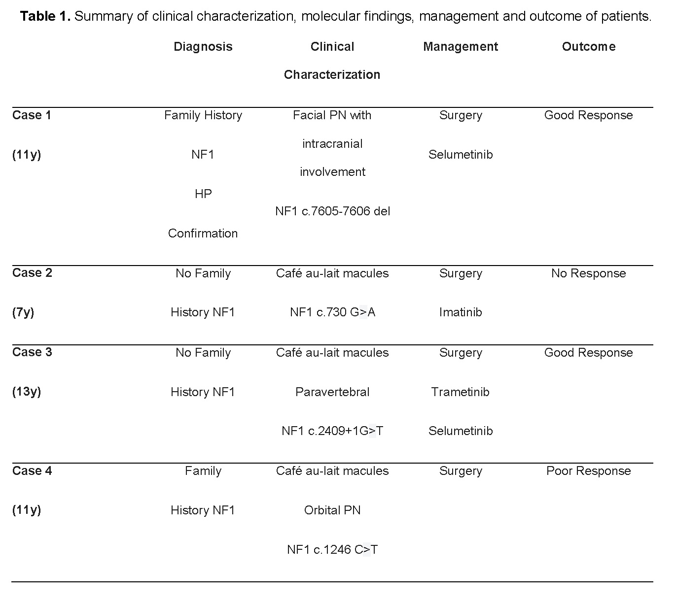
The summary of the four cases, description of the location of the lesion, pharmacological and/or surgical management and the evolution are presented in Table 1.
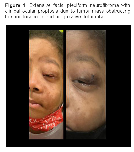
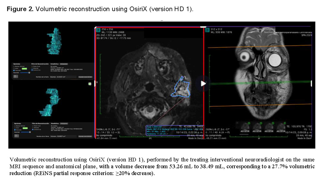
The first case involves an 11-year-old female with a family history of NF1 in a first-degree relative, confirmed by medical record review to meet NF1 diagnostic criteria. Her clinical progression began with left ocular proptosis and hearing loss due to tumor mass obstructing the auditory canal. Imaging revealed a PN involving the left orbit and cavernous sinus, progressing to involve the oral cavity. Treated with selumetinib 25 mg/m2 twice a day, she achieved significant clinical and radiological tumor reduction (27.7% of Volumetric Reduction) in 6 months (Figure 1 & 2).
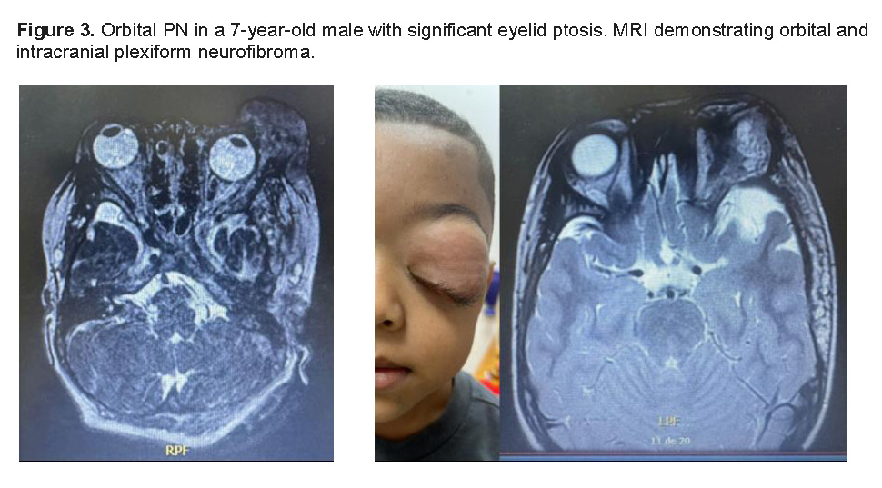
The second case describes a 7-year-old male with orbital PN and sphenoid dysplasia. Initial treatment with imatinib yielded no clinical response; the dose and duration of imatinib could not be determined, as it had been administered at another institution, and no adverse events were reported. Therapeutic failure was defined as a lack of clinical improvement in tumor size or associated symptoms. Partial surgical resections were performed approximately every six months between 2023 and 2025 without disease control, and the persistence of deformity led to the initiation of selumetinib (Figure 3).
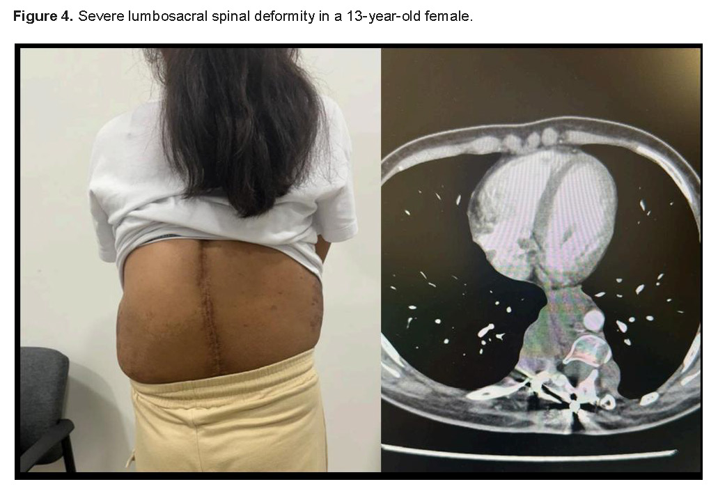
The third case is a 13-year-old female with a severe lumbosacral spinal deformity. Trametinib had been administered at another institution, where dose, duration, and adherence data were not available; therapeutic failure was defined as the absence of improvement in symptoms, size, or volume of the lesion. The documented reason for switching to selumetinib was that, at the time of presentation, selumetinib was the only MEK inhibitor with FDA approval for this indication. After spinal fixation and this failed trial of trametinib, increasing pain necessitated the initiation of selumetinib. An initial MRI was performed showing the presence of a 6.4 x 5.2 x 3.4cm paravertebral lesion at the L2-L3 level, which was no longer evident on the five-month follow-up study; both studies were compared using the same imaging protocol and anatomical plane.
Clinically, this patient showed improvement in pain, assessed with the Numeric Rating Scale (NRS) at baseline and every three months, decreasing from 7 (severe pain) to 3 (mild pain); she was not receiving concomitant analgesia that could have influenced this result. She reported an improvement in her gait, which was more agile, and an improvement in her quality of life, stating that she could sleep on her back, something she had not been able to do for 10 years (Figure 4).
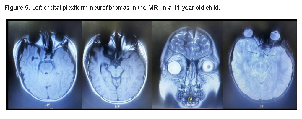
The fourth case involves an 11-year-old female with orbital PN and a family history of NF1 in a first-degree relative, confirmed by medical record review to meet NF1 diagnostic criteria. Neuropsychological evaluation using the Wechsler Intelligence Scale for Children, Fifth Edition (WISC-V), yielded a Full-Scale IQ of 69; as an isolated IQ score is not sufficient to establish a diagnosis of intellectual disability without a concurrent assessment of adaptive functioning, this finding is reported here as a cognitive profile consistent with mild intellectual impairment, pending formal adaptive-behavior evaluation (Figure 5).
DISCUSSION
The clinical diagnosis of plexiform neurofibromas (PN) remains a cornerstone of neurofibromatosis type 1 (NF1) management, as these complex tumors are often congenital and can undergo phases of rapid growth during early childhood [5,12]. Unlike superficial cutaneous neurofibromas, PNs are structurally complex, highly vascularized, and involve multiple nerve fascicles, which explains their aggressive clinical presentation. In our pediatric cohort, the heterogeneity of symptoms-ranging from progressive ocular proptosis and facial asymmetry to severe spinal deformities-highlights the clinical unpredictability of these benign but destructive lesions [2,10].
Family history plays a vital role in early identification, serving as a powerful diagnostic indicator in half of our reported cases [11]. In familial cases, the clinical suspicion of NF1 allows for proactive clinical surveillance, facilitating the discovery of deep-seated PNs before severe functional or cosmetic impairment occurs [2,3]. Conversely, in patients without a known family history, a plexiform neurofibroma frequently acts as the definitive primary clinical criterion required to confirm an NF1 diagnosis [7,13]. These apparently sporadic cases, which may arise from de novo mutations in the NF1 gene, often present a delayed diagnostic challenge until the physical mass becomes visible or symptomatic [1,8].
Radiological diagnosis, primarily achieved through high-resolution Magnetic Resonance Imaging (MRI), is essential for defining exact tumor borders, determining overall volumetric involvement, and identifying associated structural anomalies like sphenoid dysplasia [14,15]. On MRI sequences, PNs classically exhibit a hyperintense signal on T2-weighted images with a characteristic hypointense central zone, widely known in neuro-oncology as the "target sign" [16]. Longitudinal imaging follow-up is critical, as it serves as the primary method to objectively calculate changes in tumor volume, monitor growth kinetics, and evaluate therapeutic success over time [14,17].
Despite the precision of modern neuroimaging, radiological assessment in the pediatric population carries significant technical challenges, particularly because sedation may be required, depending on age, cooperation, protocol duration, and institutional resources, to avoid motion artifacts in younger children [17]. Furthermore, the infiltrative and irregular growth patterns of craniofacial and pelvic PNs complicate the manual and semi-automated segmentation of tumor borders, making volumetric tracking technically demanding [14,18]. These complexities underscore the absolute necessity of standardized imaging protocols interpreted by dedicated pediatric neuroradiologists [11,15].
The overarching therapeutic management of NF1-associated PN has evolved dramatically from purely cytoreductive surgical attempts to a specialized, multidisciplinary medical approach [11,19]. Traditionally, surgical intervention was considered the exclusive management strategy; however, total surgical resection is rarely achieved due to the tumor’s tendency to encase vital neurovascular structures and its intense intraoperative bleeding risk [20,21]. Because partial resections are frequently associated with a high rate of rapid tumor regrowth, surgical indications must be individualized and may include pain, functional deficit, disfigurement, suspicion of malignant transformation, or a reasonable prospect of resection with acceptable morbidity [12,15].
The fundamental discovery that the RAS/MAPK signaling pathway is constitutively activated in NF1 due to a loss of neurofibromin function has completely revolutionized the therapeutic framework for these patients [22,23]. This molecular mechanism provided the exact biological rationale for introducing targeted small-molecule inhibitors to disrupt the hyperactive downstream signaling pathway [23]. Consequently, MEK 1/2 inhibitors have emerged as the primary non-surgical intervention capable of altering the natural progression of progressive, inoperable, or highly symptomatic plexiform neurofibromas.
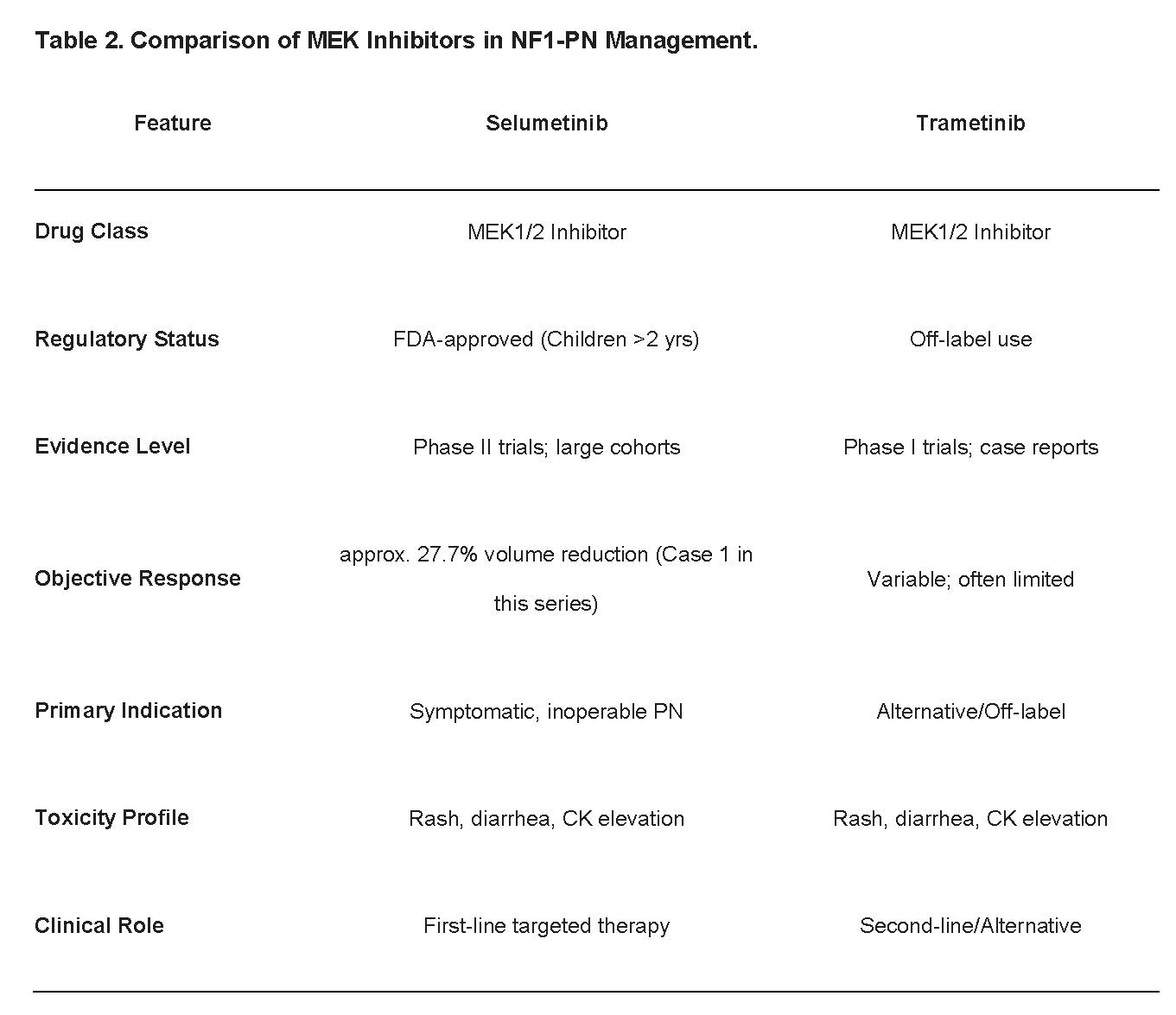
Selumetinib is currently the systemic treatment with the strongest evidence base and the broadest pediatric regulatory approval for symptomatic, inoperable NF1-associated plexiform neurofibromas (PN). This is backed by robust phase 2 clinical trial evidence demonstrating sustained tumor volume reductions of 20% or more in approximately 70% of pediatric cohorts. Furthermore, its efficacy in reducing tumor volume, chronic and acute pain, and analgesic dependency is currently being evaluated in a double-blind, phase 3 study [24,25,26].
Beyond tumor shrinkage, patients treated with selumetinib experience substantial improvement in key clinical areas, including pain reduction, motor function recovery, and overall health-related quality of life [10,24]. Real-world clinical updates continue to validate its safety, though close clinical monitoring is mandatory to manage known class-specific toxicities such as papulopustular rash, gastrointestinal distress, and asymptomatic creatine kinase elevations [27,28].
Other selective MEK inhibitors, such as trametinib and mirdametinib, are actively being utilized as crucial alternative therapies in current neuro-oncology practices [29]. These alternative agents are particularly valuable in resource-constrained regions or healthcare settings where access to selumetinib is strictly limited by regulatory approvals or high economic barriers [30]. However, as illustrated by the poor response to trametinib in one patient in our current series, interpatient variability in clinical response remains prominent, particularly given that trametinib and mirdametinib remain, in most jurisdictions, alternatives of lower evidentiary strength than selumetinib-trametinib supported mainly by phase I/II trial data and case reports and used off-label for this indication-rather than options equivalent to selumetinib; regulatory approval status differs by jurisdiction and should be verified against primary sources (FDA, EMA, or the national regulatory agency of Panama) at the time of submission, emphasizing the need for personalized treatment selection and long-term data collection [30–32] (Table 2).
CONCLUSION
Early clinical identification and expedited referral to specialized multidisciplinary centers are absolutely critical for the effective management of plexiform neurofibromas in pediatric patients with neurofibromatosis type 1. While surgical interventions remain severely limited by the highly infiltrative, hypervascular nature of these tumors and carry substantial structural risks, targeted molecular therapies using MEK inhibitors represent a profound paradigm shift in modern neuro-oncology. The remarkable clinical and radiological responses observed in our series-such as rapid volumetric tumor reduction, significant alleviation of chronic debilitating pain, and the restoration of long-lost physical functions-underscore that the universal availability and uninterrupted access to these innovative medical treatments are meaningful therapeutic options for carefully selected patients, with the potential to improve function and quality of life. These findings should be interpreted in light of the limitations of this series: a small, retrospective, single-center cohort with heterogeneous phenotypes, treatments, and follow-up, no comparator group, and possible selection bias; no inference regarding survival outcomes can be drawn from these data.
Acknowledgments
The authors wish to thank the team at the Department of Pediatrics / Oncology - Genetic Unit at Children's Hospital Dr. José Renan Esquivel for their support and the Department of Pediatrics Genetic Unit at High Complexity Pediatric Hospital of the Social Security.
Author Contributions
QK Writing: review & editing, Writing: original draft, Visualization, Project administration, Methodology, Investigation; CL: Writing: review & editing; BA: Imaging Editing; CE: Imaging Editing; SO:Writing - review & editing; CT: Writing: review & editing; HI: Writing: review & editing.
Support
Medical writing support for this article was provided by Karen Courville, MD PhD, and was funded by authors.