
INTRODUCTION
The TTN gene encodes titin, the largest known human protein and a major structural component of the sarcomere [1]. Titin spans half of the sarcomere from the Z-disk to the M-line and contributes to myocardial elasticity, passive tension, mechanosensing, and sarcomeric assembly [1,2]. Its modular architecture includes immunoglobulin-like domains, fibronectin type III domains, elastic regions, and kinase-related elements essential for cardiac muscle function [2]. Extensive alternative splicing generates multiple tissue-specific TTN isoforms, including the short Novex-3 isoform (transcript NM_133379.5, protein NP_596870.2, 5,604 amino acids), which possesses a unique, shorter C-terminal exon not shared with the larger canonical isoforms [1].
Truncating variants in TTN are among the most important genetic contributors to dilated cardiomyopathy and have also been associated with other hereditary cardiomyopathies and skeletal muscle disorders [3,4]. Nevertheless, the clinical interpretation of TTN variants remains challenging because not all truncating variants are pathogenic, and their impact depends on which transcript(s) they affect, isoform expression, protein domain localization, evolutionary conservation, population frequency, and predicted structural effect [5].
In a Panamanian cohort of patients with hereditary cardiomyopathies and arrhythmias, Cedeño-Escudero et al. reported the TTN NM_133379.5: c.11806C>T; p.(Arg3936Ter) variant in a 38-year-old female patient with dilated cardiomyopathy [6]. This nonsense variant is located in exon 46 of NM_133379.5 and affects an immunoglobulin-like region of titin. Because this alteration introduces a premature termination codon, it is predicted to generate a truncated protein and disrupt downstream structural domains.
Recent advances in artificial intelligence–based protein structure prediction, particularly AlphaFold2 and ColabFold, have created new opportunities for evaluating the structural consequences of rare variants [7,8]. These are computational, sequence-based prediction tools: used with a genuine multiple sequence alignment and cross-validated by standard structural alignment methods, they can provide meaningful, quantitative evidence regarding domain-level conservation and loss - evidence that is complementary to, but distinct in kind from, established genetic and population-level evidence.
In this study, we pursued three related aims. First, we aimed to consolidate the clinical interpretation of the TTN p.Arg3936Ter variant using established genomic evidence and ACMG/AMP interpretation. Second, we aimed to characterize the domain-level and structural consequence of the predicted truncation by combining protein domain annotation with AlphaFold2-based structural modeling of both the wild-type and truncated protein, cross-validated by quantitative structural alignment. Third, we aimed to determine the transcript specificity of this consequence across annotated TTN isoforms and its predicted nonsense-mediated decay (NMD) status, since the biological relevance of a truncating variant depends on which transcript(s) translate it.
MATERIALS AND METHODS
This study consisted of a secondary in silico characterization of the TTN NM_133379.5: c.11806C>T; p.(Arg3936Ter) variant previously identified in a Panamanian patient with dilated cardiomyopathy - the same patient and variant originally reported by Cedeño-Escudero et al. [6]. The original study was approved by the Institutional Research Ethics Committee of the Caja de Seguro Social, Panama (protocol DENSYPS-DENADOI-N-404-2024). Targeted next-generation sequencing (NGS) was performed using the SOPHiA EXTENDED CARDIO SOLUTION™ panel covering 128 genes associated with hereditary cardiomyopathies and arrhythmias on the Illumina MiniSeq platform. Variant annotation and interpretation were conducted using SOPHiA DDM® [6], Franklin by Genoox, and VarSome [7–8].
Population frequency and conservation analyses were obtained from gnomAD, dbSNP, phyloP100way, and PhastCons100way databases. Functional annotation was performed using Ensembl VEP.
Transcript-specific and NMD annotation
To determine whether the truncating consequence of this variant is shared across TTN isoforms or specific to a subset of transcripts, the variant (chr2:178,750,594 G>A, GRCh38) was annotated using Ensembl VEP against all 14 curated TTN transcripts. The genomic structure of NM_133379.5 (46 exons; ENST00000360870.10/11) was independently confirmed using the NCBI GenBank record and the Ensembl exon table, and the position of the variant within this transcript was determined by direct coordinate mapping.
Nonsense-mediated decay (NMD) susceptibility was assessed using the standard positional rule (premature termination codon outside the last exon and outside the final 50 nucleotides of the penultimate exon–exon junction), as independently reported by VarSome for NM_133379.5 specifically [8], and cross-checked against the general EJC-dependent NMD model described in the literature [17,18].
Protein domain annotation
Domain boundaries within the wild-type fragment (residues 3399–4100, 702 amino acids) were annotated using InterProScan (Pfam, SUPERFAMILY, Gene3D, SMART, and ProSite Profiles member databases). Domains were classified as immunoglobulin-like (Ig-like) of the I-set family when supported by concordant hits from at least three independent member databases (Pfam PF07679; IPR013098) [19].
Structural modeling parameters
Structural prediction of both the wild-type fragment (residues 3399–4100, 702 amino acids) and the truncated fragment (residues 3399–3935, 538 amino acids) of isoform Novex-3 (NP_596870.2) was performed using ColabFold v1.6.2 (host api.colabfold.com), implementing the AlphaFold2 pipeline in the alphafold2_ptm (monomer) configuration [9,10], with a multiple sequence alignment generated via MMseqs2 in UniRef+Environmental mode (msa_mode = mmseqs2_uniref_env) and no structural templates (template_mode = none).
Five models were generated per fragment (num_models = 5) with 3 recycles (num_recycles = 3) and 1 seed (num_seeds = 1), ranked by mean predicted local distance difference test score (rank_by = pLDDT); the top-ranked model (rank_1) for each fragment was carried forward for analysis.
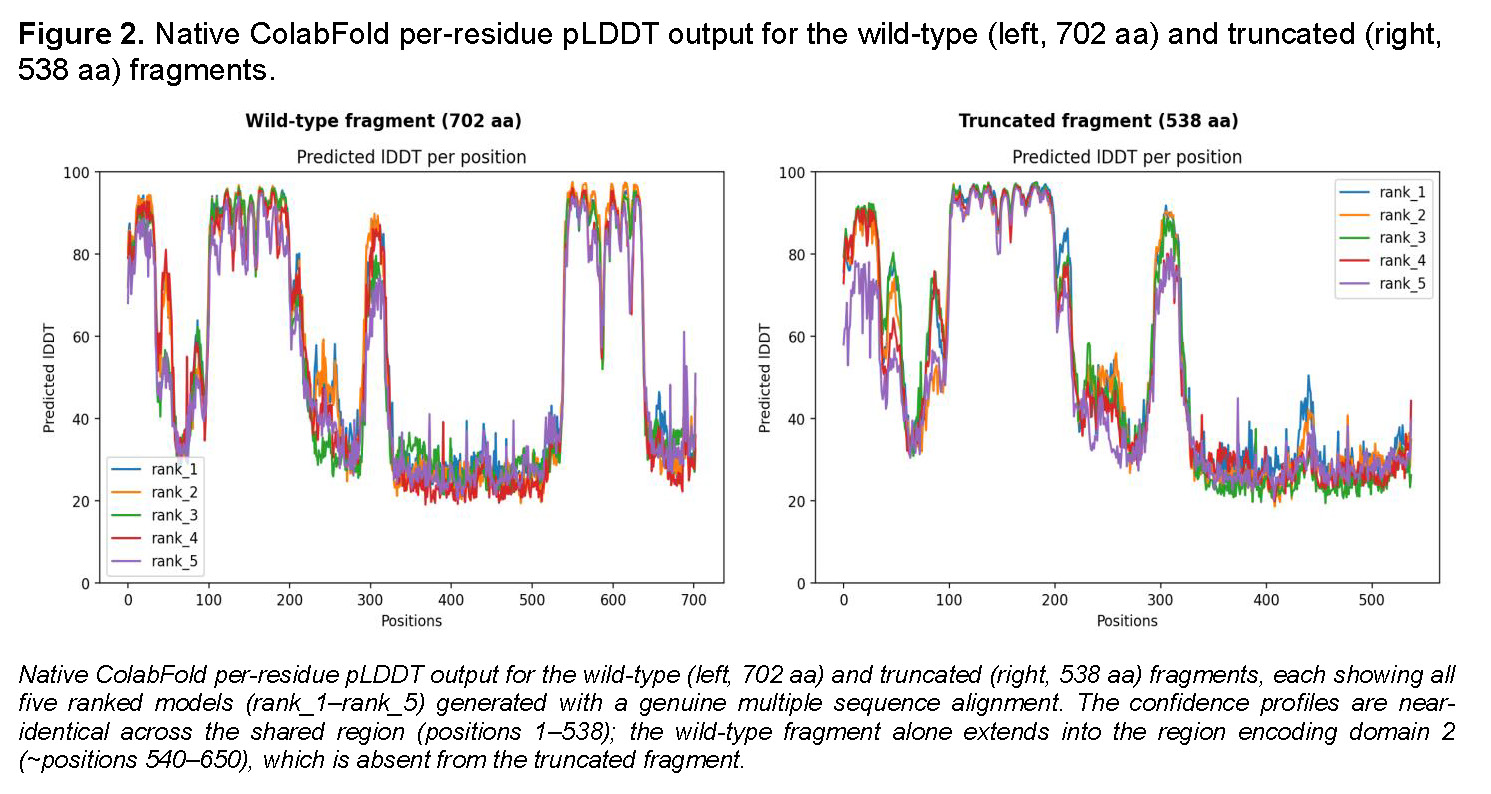
The wild-type run used a multiple sequence alignment of 5,740 sequences; the truncated run used 6,287 sequences. Predicted template modeling (pTM) scores were additionally examined for each model. For the wild-type fragment, mean pLDDT across the five ranked models was 56.5, 55.6, 53.9, 53.3, and 52.2 (rank_1–rank_5; pTM 0.30, 0.31, 0.34, 0.30, 0.31). For the truncated fragment, mean pLDDT was 55.2, 53.2, 53.1, 52.3, and 49.7 (rank_1–rank_5; pTM 0.33, 0.36, 0.37, 0.37, 0.31).
Structural comparison
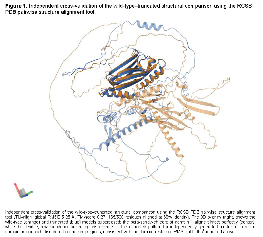
Structural similarity between the top-ranked wild-type and truncated models was assessed in two independent ways. First, Cα coordinates of the shared region (residues 1–538 of the modeled fragment) were superposed using the Kabsch rigid-body alignment algorithm, computed both globally and separately for individual well-resolved sub-regions (pLDDT > 70), to avoid the confound of independently flexible, low-confidence linker regions dominating a single global fit. Second, the full-length wild-type and truncated models were independently cross-validated using the RCSB PDB pairwise structure alignment tool (TM-align algorithm) [20].
RESULTS
Genetic and population-level evidence for pathogenicity
The TTN c.11806C>T, p.(Arg3936Ter) variant identified in the Panamanian patient with dilated cardiomyopathy corresponds to a heterozygous nonsense variant affecting the immunoglobulin-like (Ig-like) region of titin. This single nucleotide substitution introduces a premature termination codon at amino acid position 3936, replacing arginine with a stop codon and generating a truncated protein predicted to truncate approximately 29.8% of the Novex-3 isoform sequence. The variant was classified as Likely Pathogenic according to ACMG/AMP criteria by both VarSome (9 points: PVS1 very strong + PM2 moderate) and Franklin, mainly supported by PVS1 (very strong evidence for loss-of-function variants in a gene where LoF is an established disease mechanism) and PM2 (absence or extremely low frequency in population databases). Population analysis in gnomAD [11] showed a very low allele frequency in both exomes and genomes, with no reported homozygous individuals. Conservation analysis demonstrated that the affected residue is evolutionarily constrained, with high phyloP100way (4.024) and PhastCons100way (0.943) scores. Additional in silico prediction revealed a CADD score of 15.4, while SpliceAI and Pangolin analyses showed no significant splice alteration effects, consistent with protein truncation rather than aberrant splicing as the predominant sequence-level mechanism. The transcript-specific, domain-level, and structural consequences of the truncation are examined in the following subsections.
Transcript specificity and NMD status
Ensembl VEP annotation of chr2:178,750,594 G>A across the 14 curated TTN transcripts showed that this genomic position is annotated as an intronic variant in 91% of transcripts, produces a downstream-of-gene-body consequence in one transcript lacking this region, and generates a stop-gained consequence exclusively in NM_133379.5 (Novex-3) - the transcript on which the original clinical variant nomenclature is based. Independent examination of the NM_133379.5 exon structure (46 exons total, confirmed via the NCBI GenBank record and the Ensembl exon table) showed that the variant falls within exon 46 - the final exon of this transcript, at position 1,446 of a 7,635-nucleotide terminal exon. Consistent with the classical exon-junction-complex (EJC) model of NMD, in which a premature termination codon located in the last exon lacks a downstream EJC and is therefore not expected to trigger decay [17,18], VarSome’s automated PVS1 evidence for NM_133379.5 explicitly states that this variant is not predicted to cause NMD. This differs from an initial assessment by Franklin, which predicted NMD based on the variant’s position within the canonical, 362-exon meta-transcript of TTN - a transcript in which this position is not the terminal exon; Franklin’s own annotation explicitly identifies NM_133379.5 as the “non-canonical” transcript for this evaluation. This discrepancy illustrates the importance of evaluating NMD status against the specific transcript relevant to a given clinical variant, rather than against a gene’s canonical or meta-transcript by default.
Domain-level consequence of the truncation
InterProScan annotation of the 702-residue wild-type fragment (residues 3399–4100) identified two immunoglobulin-like (Ig-like) domains of the I-set family (Pfam PF07679; IPR013098), independently supported by Pfam, SUPERFAMILY, Gene3D, SMART, and ProSite Profiles: domain 1 spanning fragment positions 98–205, and domain 2 spanning fragment positions 540–646, separated by an unannotated linker (positions ~232–539) containing a short coiled-coil segment (positions 211–231). The p.Arg3936Ter variant corresponds to fragment position 538 - three residues upstream of the annotated start of domain 2. Because the truncated protein terminates at fragment position 537, it retains domain 1 in full but loses domain 2 in its entirety (0 of 107 residues retained).
Computational structural modeling and structural alignment
Both the wild-type (702-residue) and truncated (538-residue) fragments were modeled using ColabFold with a genuine multiple sequence alignment (see Methods). The top-ranked wild-type model achieved a mean pLDDT of 56.5 (pTM = 0.30); the top-ranked truncated model achieved a nearly identical mean pLDDT of 55.2 (pTM = 0.33). Per-residue pLDDT profiles for the two models were closely superimposable across the entire shared region (residues 1–538), with confident peaks (pLDDT > 70) corresponding to domain 1 (positions 98–205) and two additional well-resolved sub-regions, separated by lower-confidence linker stretches, in both models.
Quantitative structural superposition confirmed this visual similarity. A single global rigid-body superposition of the full shared region (538 residues) yielded a large RMSD (>40 Å), reflecting the fact that independently modeled, low-confidence flexible linkers adopt divergent relative orientations between separately generated models - a well-recognized artifact of whole-chain superposition in multi-domain proteins with disordered linkers, rather than evidence of true structural divergence. Restricting superposition to individual well-resolved sub-regions eliminated this artifact: domain 1 alone (fragment positions 98–205, 108 residues) superposed between wild-type and truncated models with an RMSD of 0.19 Å, indicating an essentially identical fold. Independent cross-validation using the RCSB PDB pairwise structure alignment tool (TM-align) on the full-length models returned a global RMSD of 5.25 Å and TM-score of 0.27 (165 of 538 residues aligned with confidence, 69% sequence identity in the aligned region) - consistent with the same interpretation: the well-folded core regions align closely (visually confirmed in the RCSB structural overlay, where the beta-sandwich core of domain 1 superposes almost perfectly), while the global metric is diluted by the flexible, independently-modeled linker regions. Because the truncated fragment terminates at position 537, it altogether lacks the sequence encoding domain 2 (positions 540–646); this is not a modeling artifact but a direct, necessary consequence of the truncation, independently confirmed by the InterProScan domain annotation above.
DISCUSSION
Truncating variants in TTN (TTNtv) are recognized as the most common genetic cause of dilated cardiomyopathy (DCM), accounting for approximately 10–25% of familial and idiopathic cases worldwide [1,3]. In the present study, we characterized the nonsense variant TTN c.11806C>T; p.(Arg3936Ter), identified in a Panamanian patient with DCM. This variant introduces a premature termination codon predicted to truncate the Novex-3 isoform of titin after residue 3936, removing a complete C-terminal immunoglobulin-like domain.
The affected residue demonstrated strong evolutionary conservation, supported by elevated PhastCons and PhyloP scores, and the variant showed an absence or extremely low frequency in population databases, fulfilling ACMG/AMP criteria PVS1 and PM2 for pathogenicity classification. Similar findings have been reported in previous studies demonstrating that TTN truncating variants affecting cardiac-relevant exons are enriched in DCM patients and are associated with more severe ventricular dysfunction [12]. Furthermore, León et al. described that splice-disrupting and truncating variants in TTN contribute significantly to familial DCM, emphasizing the importance of comprehensive molecular characterization in inherited cardio- myopathies [16].
Recent studies have emphasized the importance of integrating structural and functional evidence, and of transcript-level context, for the interpretation of TTN variants [14,15]. Artificial intelligence–based structural prediction tools such as AlphaFold2 have expanded the capacity to evaluate the potential structural consequences of rare genomic variants [9,10]. In our analysis, standard domain annotation (InterProScan/Pfam) established that the p.Arg3936Ter truncation eliminates a complete C-terminal Ig-like domain, and MSA-based AlphaFold2 modeling of both the wild-type and truncated fragments - combined with quantitative structural alignment (Figure 1) - independently confirmed that the retained portion of the protein folds identically (RMSD 0.19 Å for the retained domain) while the second domain is simply absent from the truncated model, exactly as predicted by domain annotation. This convergence between an annotation-based method and an independent, quantitative structural method (across two alignment approaches) provides strong, mutually reinforcing evidence for the structural consequence of this variant.
Transcript-specific interpretation of TTN truncating variants
A central finding of this study is that the truncating consequence of p.Arg3936Ter is not shared across TTN isoforms: VEP annotation showed a stop-gained consequence exclusively in NM_133379.5 (Novex-3), while the same genomic position is intronic in 91% of annotated transcripts. AlphaFold2 modeling of both fragments (Figure 2) confirms that this is a clean loss of the terminal domain rather than a general destabilization of the protein. This is expected given that NM_133379.5 possesses a unique, short C-terminal exon not present in the canonical isoforms [1], but it underscores that the clinical relevance of this variant depends specifically on expression and translation of the Novex-3 isoform - the isoform on which the original clinical report and variant nomenclature are based [6]. This finding also resolves an internal inconsistency between automated tools: Franklin’s NMD prediction, based on the canonical meta-transcript, would misclassify this variant’s NMD status if applied without reference to the specific clinical transcript; VarSome’s transcript-specific evaluation, and our independent confirmation via the NM_133379.5 exon structure and standard EJC rules, converge on the correct conclusion that this variant escapes NMD and is expected to yield a stable truncated protein.
This has a direct mechanistic implication: because the truncated transcript escapes NMD, the truncated protein product is expected to be translated and to accumulate, rather than being degraded. Combined with the domain-level and structural evidence above - complete, confirmed loss of a C-terminal Ig-like domain with an otherwise identically folded retained region - this supports a loss-of-function mechanism operating through production of a stable, domain-deficient titin protein specific to the Novex-3 isoform, rather than through haploinsufficiency via transcript degradation.
Finally, this study highlights the growing utility of structural bioinformatics and artificial intelligence–based modeling in precision cardiovascular genomics, when applied with attention to transcript-specific context and cross-validated with independent structural alignment methods. Experimental validation remains necessary to confirm the exact functional consequences of this variant; the present findings support the role of integrative computational genomics as a complementary tool in refining the interpretation of rare TTN truncating variants, particularly in underrepresented populations from Latin America.
Limitations
This study has four main limitations. First, no functional validation experiments were performed; the biological consequences described here remain computationally inferred.
Second, segregation analysis was not available to confirm co-segregation with disease.
Third, the relative cardiac expression of the Novex-3 isoform was not directly quantified, which would strengthen the interpretation of clinical relevance. Fourth, structural modeling and alignment, though cross-validated by two independent methods, remain computational predictions rather than experimentally determined structures.
CONCLUSIONS
This study supports a Likely Pathogenic, loss-of-function classification for the TTN p.Arg3936Ter variant (PVS1, PM2), identified in the same patient originally reported by Cedeño-Escudero et al. [6]. Its truncating effect is specific to the Novex-3 isoform (NM_133379.5), escapes nonsense-mediated decay, and - confirmed independently by domain annotation and cross-validated structural alignment - yields a stable protein that retains a correctly folded first Ig-like domain (RMSD 0.19 Å vs. wild-type) but completely lacks the second.
These findings illustrate the value of transcript-specific, structurally cross-validated computational analysis for interpreting rare TTN truncating variants in underrepresented populations such as Panama; functional validation and segregation studies remain necessary to confirm the proposed mechanism.
Author Contributions
Lydier De Gracia: Conceptualization, data analysis, structural modeling, manuscript drafting, review, and writing. José Cedeño: Conceptualization, supervision, manuscript review, and writing support. Omar Espinosa: Conceptualization and manuscript review. Luis Sotillo: Conceptualization, manuscript review, and scientific supervision.
Acknowledgements
The authors thank the Specialized Center for Medical Genetics and Genomics of the Social Security Fund of Panama for technical and institutional support.
Funding
This study received no external funding.